


 Foundational characteristics of cancer include proliferation, angiogenesis, migration, evasion of apoptosis, and cellular immortality. Find key markers for these cellular processes and antibodies to detect them.
 The SUMOplot™ Analysis Program predicts and scores sumoylation sites in your protein. SUMOylation is a post-translational modification involved in various cellular processes, such as nuclear-cytosolic transport, transcriptional regulation, apoptosis, protein stability, response to stress, and progression through the cell cycle.
 The Autophagy Receptor Motif Plotter predicts and scores autophagy receptor binding sites in your protein. Identifying proteins connected to this pathway is critical to understanding the role of autophagy in physiological as well as pathological processes such as development, differentiation, neurodegenerative diseases, stress, infection, and cancer.
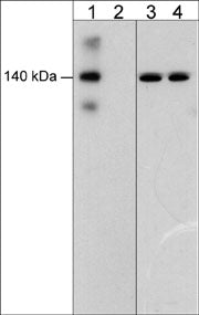
Anti-VE-Cadherin (Tyr-685), Phosphospecific Antibody
- SPECIFICATION
- CITATIONS
- PROTOCOLS
- BACKGROUND

| Primary Accession | P33151 |
|---|---|
| Reactivity | Bovine |
| Host | Rabbit |
| Clonality | Rabbit Polyclonal |
| Isotype | IgG |
| Calculated MW | 87528 Da |
| Gene ID | 1003 |
|---|---|
| Other Names | Cadherin-5, vascular endothelial Cadherin, CD144 |
| Target/Specificity | Cadherins are transmembrane glycoproteins vital in calcium-dependent cell-cell adhesion during tissue differentiation. Cadherins cluster to form foci of homophilic binding units. A key determinant to the strength of the cadherin-mediated adhesion may be by the juxtamembrane region in cadherins. VE-cadherin (Cadherin 5) is the major cadherin found in endothelial cells and has important roles during angiogenesis and maintenance of barrier permeability. The cytoplasmic domain of VE-cadherin comprises the juxtamembrane domain that binds to the p120 catenin, and the carboxylterminal domain that interacts with β- or γ-catenins. Modulation of tyrosine phosphorylation on one or more of the nine tyrosine sites in the cytoplasmic domain may be important for regulating both angiogenesis and permeability. Phosphorylation of Tyr-658 and Tyr-731 alters catenin binding, restores cell migration, and decreases barrier permeability. While VEGF-induced phosphorylation of Tyr-685 occurs through c-Src, and regulates endothelial cell migration, but not permeability |
| Format | Antigen Affinity Purified |
| Storage | Maintain refrigerated at 2-8°C for up to 6 months. For long term storage store at -20°C in small aliquots to prevent freeze-thaw cycles. |
| Precautions | Anti-VE-Cadherin (Tyr-685), Phosphospecific Antibody is for research use only and not for use in diagnostic or therapeutic procedures. |
| Shipping | Blue Ice |

Thousands of laboratories across the world have published research that depended on the performance of antibodies from Abcepta to advance their research. Check out links to articles that cite our products in major peer-reviewed journals, organized by research category.
info@abcepta.com, and receive a free "I Love Antibodies" mug.

Provided below are standard protocols that you may find useful for product applications.
Background
Cadherins are transmembrane glycoproteins vital in calcium-dependent cell-cell adhesion during tissue differentiation. Cadherins cluster to form foci of homophilic binding units. A key determinant to the strength of the cadherin-mediated adhesion may be by the juxtamembrane region in cadherins. VE-cadherin (Cadherin 5) is the major cadherin found in endothelial cells and has important roles during angiogenesis and maintenance of barrier permeability. The cytoplasmic domain of VE-cadherin comprises the juxtamembrane domain that binds to the p120 catenin, and the carboxylterminal domain that interacts with β- or γ-catenins. Modulation of tyrosine phosphorylation on one or more of the nine tyrosine sites in the cytoplasmic domain may be important for regulating both angiogenesis and permeability. Phosphorylation of Tyr-658 and Tyr-731 alters catenin binding, restores cell migration, and decreases barrier permeability. While VEGF-induced phosphorylation of Tyr-685 occurs through c-Src, and regulates endothelial cell migration, but not permeability
If you have used an Abcepta product and would like to share how it has performed, please click on the "Submit Review" button and provide the requested information. Our staff will examine and post your review and contact you if needed.
If you have any additional inquiries please email technical services at tech@abcepta.com.
Ordering Information
Other Products
Shipping Information
